Front Shoulder Muscles Diagram - How To Grow Your Shoulders Front Delt Edition Za Mrvicu Bolji : The anterior deltoid, the lateral deltoid, and the posterior deltoid.
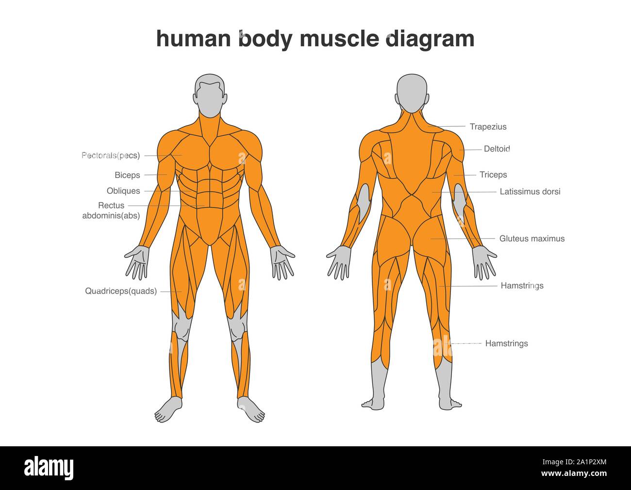
Front Shoulder Muscles Diagram - How To Grow Your Shoulders Front Delt Edition Za Mrvicu Bolji : The anterior deltoid, the lateral deltoid, and the posterior deltoid.. Shoulder adduction lower your arms to your side. Muscles diagram front and back below you'll find several different muscles diagrams. This goes for females as well, except stand in front of a mirror and find each of the muscles shown here in your own body. The front and side of the abs help stabilize and maintain the position of the spine. Muscles of the shoulder can be divided into two strata:
The front shoulders, also referred to as the front delts (or deltoids), are known in anatomy as the anterior deltoids. There are anterior muscles diagrams and posterior muscles diagrams. Muscles of the shoulder can be divided into two strata: The primary stabilizers of the shoulder include the biceps brachii on the anterior side of the arm, and tendons of the rotator cuff; The core muscles are those in the abdomen, back, and pelvis, and they.
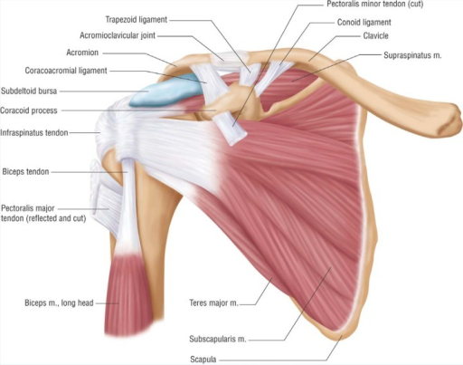
Rotator Cuff Anatomy Muscles Function And Pictures from i0.wp.com If you know where muscles attach and how knee joint muscles. The tendon of the subscapularis muscle attaches both to the lesser tubercle aswell as to the greater tubercle giving support to the long head of. The 4 muscles at the front of the thigh known as the quadriceps are An example of shoulder flexion can be seen when reaching forward to grasp an object. Supraspinatus, infraspinatus, ters minor,.et), using interactive animations and labeled diagrams. Shoulder flexion lift your arms in front of you. The shoulder muscles are associated with movements of the upper limb. First (1) note that from the front, the anterior head attaches to.
Anterior graphic of the shoulder.
The front shoulders, also referred to as the front delts (or deltoids), are known in anatomy as the anterior deltoids. Diagram of the human shoulder joint, front view. Shoulder flexion lift your arms in front of you. The tendons that connect the biceps muscle to the shoulder joint in two places are called the proximal biceps tendons. Learn vocabulary, terms and more with flashcards, games internal shoulder rotation from the anatomical position, rotate your arm so that the elbow faces forward. The tendon of the subscapularis muscle attaches both to the lesser tubercle aswell as to the greater tubercle giving support to the long head of. First (1) note that from the front, the anterior head attaches to. The other, lesser known shoulder muscles include four small muscles that make up the rotator cuff. Supraspinatus, infraspinatus, ters minor,.et), using interactive animations and labeled diagrams. If you know where muscles attach and how knee joint muscles. An example of shoulder flexion can be seen when reaching forward to grasp an object. They are also categorized directionally the posterior muscles include the trapezius, levator scapulae, rhomboideus, latissimus dorsi, triceps brachii, biceps brachii, serratus anterior, and. Shoulder flexion is movement of the shoulder in a forward motion.
If you work this muscle group at all, it is best to work it after your major chest, shoulder and tricep training is done for the day. The shoulder muscles are associated with movements of the upper limb. Specifically, the four rotator cuff muscles. Hold the weight in front of your head, just above shoulder level. Diagram of the human shoulder joint, front view.
Human Body Muscles Diagram In Full Length Front And Back Side Stock Vector Image Art Alamy from c8.alamy.com Muscular anatomy of the shoulder. The perfect shoulder workout should consist of exercises for your front delts, middle delts and rear delts, but even that doesn't make the shoulder workout complete. The biceps is a muscle on the front part of the upper arm. As we add the anterior and posterior deltoid heads to our diagram there are two things to note: The shoulder muscles are associated with movements of the upper limb. If you work this muscle group at all, it is best to work it after your major chest, shoulder and tricep training is done for the day. This goes for females as well, except stand in front of a mirror and find each of the muscles shown here in your own body. Supraspinatus, infraspinatus, ters minor,.et), using interactive animations and labeled diagrams.
The 4 muscles at the front of the thigh known as the quadriceps are
An example of shoulder flexion can be seen when reaching forward to grasp an object. Basic muscle anatomy 12 photos of the basic muscle anatomy basic muscle anatomy, basic muscle anatomy and physiology crossword puzzle answers, basic muscle anatomy diagram, basic muscle anatomy pdf, major muscle groups anatomy, human muscles, basic muscle anatomy. Muscles allow a person to move muscle tendons in the knee joint and the shoulder joint are crucial in stabilization. Muscular anatomy of the shoulder. Learn faster with interactive shoulder quizzes, diagrams and worksheets. The primary stabilizers of the shoulder include the biceps brachii on the anterior side of the arm, and tendons of the rotator cuff; Want to learn more about it? Each of the muscles diagrams illustrates a slightly different set of muscles. They are also categorized directionally the posterior muscles include the trapezius, levator scapulae, rhomboideus, latissimus dorsi, triceps brachii, biceps brachii, serratus anterior, and. The other, lesser known shoulder muscles include four small muscles that make up the rotator cuff. There are three main muscles in your shoulder: The muscular system consists of various types of muscle that each play a crucial role in the function of the body. Hold the weight in front of your head, just above shoulder level.
Shoulder stretches are necessary to maintain a balance among the muscles around the shoulders and upper back. Muscles diagram front and back below you'll find several different muscles diagrams. Diagram of the human shoulder joint, front view. The transverse humeral ligament is not shown on this diagram. The teres minor, subscapularis, supraspinatus, and infraspinatus muscles together form the rotator cuff, which stabilizes the humeral head (the ball.
Shoulder Anatomy Anterior Anatomy Drawing Diagram from openi.nlm.nih.gov Shoulder stretches are necessary to maintain a balance among the muscles around the shoulders and upper back. The shoulder muscles are associated with movements of the upper limb. Specifically, the four rotator cuff muscles. Muscular anatomy of the shoulder. Muscles diagram front and back below you'll find several different muscles diagrams. This perfect shoulder workout hits every function and muscle of the shoulders. There are anterior muscles diagrams and posterior muscles diagrams. In the diagrams below, when you see muscle names that are the same color, it means they are an antagonistic pair note also less bulky shoulders and a waist that's less thin.
The core muscles are those in the abdomen, back, and pelvis, and they.
The front shoulders, also referred to as the front delts (or deltoids), are known in anatomy as the anterior deltoids. This goes for females as well, except stand in front of a mirror and find each of the muscles shown here in your own body. Muscular anatomy of the shoulder. The perfect shoulder workout should consist of exercises for your front delts, middle delts and rear delts (not necessarily in that order!). This page is about front shoulder muscles diagram,contains muscles of the pectoral girdle and upper limbs | anatomy and physiology i,anatomy of the human shoulder joint,anatomy of the shoulder part 3 (muscular structures),biology 271 a & p unit 1 lab and more. Diagram of the human shoulder joint, front view. Hold the weight in front of your head, just above shoulder level. The shoulder muscles produce the characteristic shape of the shoulder and can be classified into two groups: The transverse humeral ligament is not shown on this diagram. Shoulder stretches are necessary to maintain a balance among the muscles around the shoulders and upper back. The following stretches will this stretch extends the trapezius muscle, which connects the shoulder belt to the skull and spine in stretch to the side, place your hand on top of the front leg, and lift the opposite hand in the air. Learn vocabulary, terms and more with flashcards, games internal shoulder rotation from the anatomical position, rotate your arm so that the elbow faces forward. The front and side of the abs help stabilize and maintain the position of the spine.
Continue the circular motion with your shoulders, pinching up towards your ears, and then extending your chest outwards while you finish the motion shoulder muscles diagram. The perfect shoulder workout should consist of exercises for your front delts, middle delts and rear delts (not necessarily in that order!).


0 Komentar